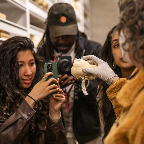
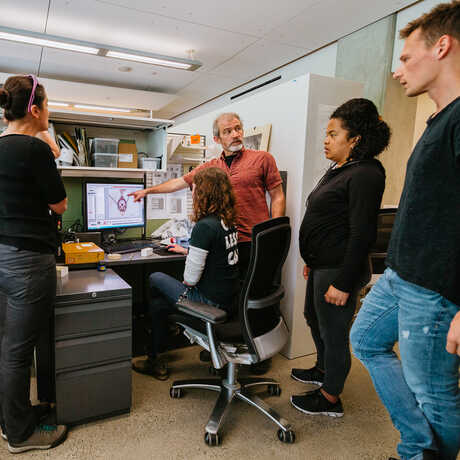
The mission of the Academy's Institute for Biodiversity Science and Sustainability (IBSS) is to gather new knowledge about life's diversity and the process of evolution—and to rapidly apply that understanding to our efforts to regenerate life on Earth.
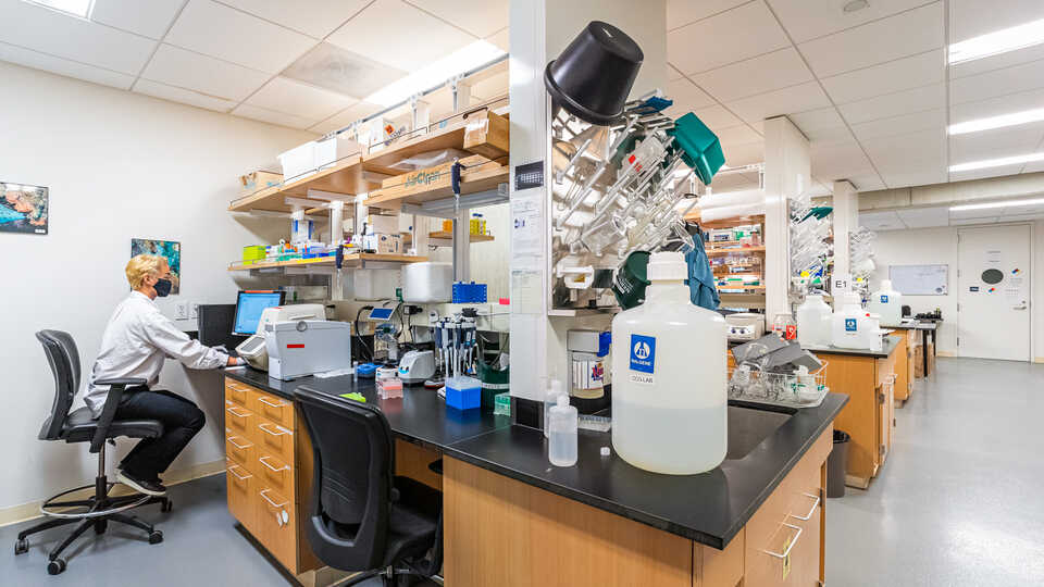
The Academy's Institute for Biodiversity Science and Sustainability offers a number of resources to support practicing scientists in their research efforts, as well as opportunities for students looking for mentorship and hands-on experience in scientific research.
Resources for Practicing Scientists
Center for Comparative Genomics
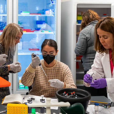
The Center for Comparative Genomics (CCG) serves as the Academy's central hub for genomics and sequencing. Our facilities include a fully-equipped genomics laboratory, a frozen DNA CryoCollection, and high-performance computing resources. The CCG focuses on museum genomics, employing the latest molecular and computational techniques and utilizing natural history specimens to explore questions relevant to biodiversity, conservation, and regeneration. Our primary mission is to spearhead groundbreaking research in museum genomics at the Academy. The CCG engages in diverse projects spanning the breadth of the tree of life and spanning the globe—From the Caribbean coral reefscapes and Galápagos birds to frogs of Cameroon, California native plants, and bioluminescent fish microbiomes in the Indo-Pacific. In addition to our research efforts, we are dedicated to training and supporting the next generation of biodiversity researchers. As leaders in museum genomics, the CCG hosts workshops and training sessions for local researchers interested in the field. Our aim is to create a supportive environment for scientific exploration and knowledge sharing.
More about the CCG
More about CCG Projects
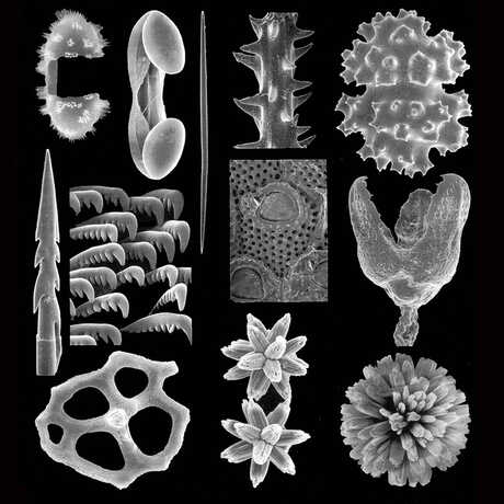
Scanning Electron Microscopy Lab
The Scanning Electron Microscopy (SEM) lab consists of a LEO/Zeiss 1450 VP SEM and several pieces of ancillary equipment. The SEM is based on a tungsten thermionic electron gun and is capable of magnifications above 100,000X. Features as small as 50 nanometers can be resolved. The SEM images are in digital TIF format, with a maximum resolution of 3072x2300 pixels. Normally all specimens must be fully dried and mounted on an SEM stub and coated with a very thin layer of gold before being put in the SEM. The instrument has 3 electron detectors: a normal secondary electron (SE) detector, a variable pressure electron detector, and a backscatter detector. There is also a vacuum sputter coater for coating specimens with a thin layer of gold (around 20nm thickness) and a CO² critical point drier. The lab is located in a new space designed specifically for the SEM on the lowest level of the building so as to eliminate vibrations that would otherwise ruin the imaging. The lab is supervised by Scott Serata and I am available to either run the SEM for researchers or train users so they can run by themselves.
Opportunities for Practicing Scientists
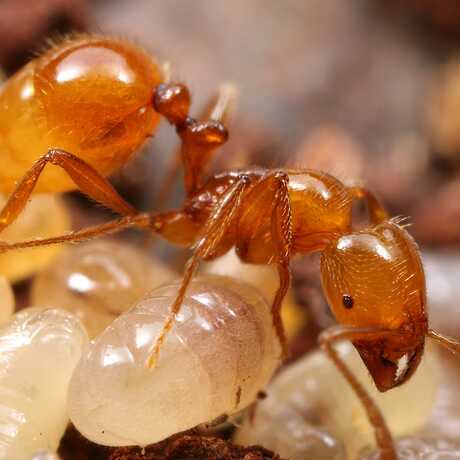
Ant Course
Ant Course is a workshop designed primarily for systematists, ecologists, behaviorists, conservation biologists, and other biologists whose research responsibilities require a greater understanding of ant taxonomy and field research techniques. Emphasis is on the evolution, classification, and identification of ant genera. Lectures include background information on the ecology, life histories, and evolution of ants. Field trips emphasize collecting and sampling techniques, and associated lab work focuses specimen preparation, sorting, and labeling. Information on equipment, literature, and myrmecological contacts are also presented.
Learn more
Opportunities for Students
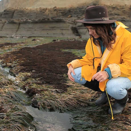
Summer Systematics Institute
Gain hands-on experience while working alongside renowned scientists—and jump-start your career in natural sciences research—with an eight-week, museum-based, paid internship. SSI offers undergraduates important insights into the contributions that collections-based research can make to critical issues such as worldwide threats to biodiversity, and the origins and diversification of life.
Learn more
Biological Illustration Internship
Collaborate with Academy scientists to develop publication-ready scientific illustrations of biological specimens during an eight-week, paid summer internship. Using ink, watercolor, and/or computer-assisted design, the Biological Illustration intern will learn how to present clear, accurate visual messages about scientific subject matter.
Learn more
Graduate Student Advisors
The Institute for Biodiversity Science and Sustainability and San Francisco State University have a long standing partnership and many of the Curators at the California Academy of Sciences have graduate faculty status in the Biology Department as Research Professors. As faculty of both the California Academy of Sciences and San Francisco State University, they may accept and direct their own graduate students. Graduate students advised by Academy Curators/Research Professors are enrolled at SFSU in the Biology Department. To learn more about the opportunity for applying to SFSU, under the advisory of a Curator/Research Professor from the California Academy of Sciences, please visit https://biology.sfsu.edu/graduate
Current Academy Research Professors are as follows:
Rebecca Albright, Assistant Curator, Invertebrate Zoology
Shannon Bennett, Chief of Science, Hind Dean of Science and Research Collections, Associate Curator, Microbiology
Jack Dumbacher, Curator, Ornithology and Mammalogy
Lauren Esposito, Assistant Curator and Schlinger Chair of Arachnology
Brian Fisher, Patterson Scholar and Curator, Entomology
Terry Gosliner, Senior Curator, Invertebrate Zoology
Sarah Jacobs, Assistant Curator and Howell Chair of Western North American Botany
Rebecca Johnson, Co-Director, Community Science and Research Associate, Invertebrate Zoology
Durrell Kapan, Senior Research Fellow, Entomology
Rich Mooi, Curator, Invertebrate Zoology
Luiz Rocha, Associate Curator and Follett Chair of Ichthyology
Peter Roopnarine, Curator, Invertebrate Zoology and Geology
Brian Simison, Associate Curator and Director of the Center for Comparative Genomics
Michelle Trautwein, Assistant Curator and Schlinger Chair of Diptera
Gary Williams, Curator of Invertebrate Zoology
Prospective volunteers, we offer a wide range of opportunities to match nearly any interest, whether your preference is helping visitors to explore our exhibits, preparing specimens in our research collections, or caring for our aquarium tanks.
